The timing of a pregnancy ultrasound depends on the clinical question: confirming an early pregnancy, screening for chromosome-risk markers, examining fetal anatomy, or monitoring growth and wellbeing. By the end, you will know which scans are commonly scheduled, which are ordered only for a specific concern, how to prepare, and when to seek prompt assessment.
Key takeaways
- Seek an early scan for uncertain dates, bleeding, pain, or ectopic pregnancy concerns.
- Schedule the nuchal translucency scan from 11 weeks to 13 weeks 6 days.
- The anatomy scan at 18–22 weeks examines organs, placenta, fluid and fetal development.
- Later scans assess growth, fluid, Doppler blood flow, movement and presentation.
Early pregnancy scans: confirming location, heartbeat, dates, and number of fetuses
An early ultrasound is needed when dates are uncertain, bleeding or pelvic or abdominal pain occurs, or examination raises concern about ectopic pregnancy or miscarriage. It may also follow a previous pregnancy complication or provide a clinically indicated first-trimester assessment.
It can establish whether the pregnancy is inside the uterus, show fetal heartbeat when visible at that stage, estimate gestational age, identify the number of fetuses, and assess the cervix, uterus, ovaries, or other pelvic structures.
| Examination | How it is performed | When it is useful |
|---|---|---|
| Transvaginal ultrasound | A covered probe is placed in the vagina for closer early images | Uncertain dates, pain, bleeding, suspected ectopic pregnancy or miscarriage, and close assessment of the cervix or pelvis |
| Abdominal ultrasound | Gel is applied to the abdomen and a probe moves over the skin | When abdominal views provide sufficient detail or a wider overview is needed |
A transvaginal scan does not mean the pregnancy is abnormal; it is often the clearest early examination. It usually requires an empty bladder. An abdominal scan may require a comfortably full bladder, so follow the imaging centre’s instructions.
Weeks calculated from the last menstrual period can differ from ultrasound dating, especially with irregular cycles or late ovulation. An early clinician-assessed scan is more useful for dating than a later scan because fetal growth becomes less precise for assigning the original gestational age.
Search results for ultrasound during pregnancy in Navi Mumbai should therefore be matched to the clinical question, not treated as interchangeable scans.
The 11-week to 13-week 6-day scan: nuchal translucency and early screening
The nuchal translucency scan is performed from 11 weeks 0 days to 13 weeks 6 days, when the crown-rump length measures 45–84 mm. It measures the fluid space at the back of the fetal neck to help assess chromosome risk. It is a screening measurement, not a diagnosis.
A planned first-trimester scan also checks the fetal heartbeat, confirms dating, reviews early anatomy, and counts fetuses. In twins or higher-order pregnancies, it should document chorionicity and amnionicity: the number of placentas and amniotic sacs. These findings affect later surveillance.
The scan usually assesses:
- Whether the nuchal translucency measurement is technically valid and how it fits the clinician’s chromosome-screening plan.
- Fetal size, heartbeat, dating, early anatomy, and the number of fetuses.
- In a multiple pregnancy, which fetuses share a placenta or amniotic sac.
This scan is not automatically required for every pregnancy. Your clinician may offer it as part of first-trimester screening, but the decision depends on your overall screening plan, clinical circumstances, and informed choice. A measurement requiring follow-up does not confirm a chromosome condition; it indicates that additional assessment may be appropriate.
Do not confuse it with an urgent scan for pain or bleeding. That examination answers immediate safety questions, such as pregnancy location and viability, rather than serving as a planned nuchal translucency appointment. When arranging a pregnancy ultrasound in Airoli, ask how the report will be integrated with your obstetrician’s plan.
The 18–22 week anatomy scan: checking organs, placenta, and fluid
The 18–22-week anatomy scan asks whether the fetus is developing normally in structure, and whether the placenta and amniotic fluid appear appropriate for this stage.
- The sonographer reviews the brain, face, spine, chest, heart, abdomen, kidneys, limbs, and fetal measurements.
- The examination records placenta location, including whether it is low-lying, and assesses amniotic-fluid volume.
- The report states the indication, gestational age, findings, examination limitations, and recommended next step.
This planned scan suits an uncomplicated pregnancy, but timing changes when an earlier result, symptoms, multiple pregnancy, or a maternal condition requires specialist follow-up.
A scan can identify many structural abnormalities, but it cannot exclude every birth defect, genetic condition, heart problem, or developmental issue. A normal result is reassuring, not a guarantee that every condition is absent.
A limited view does not automatically mean something is wrong. Fetal position, gestational age, maternal body habitus, equipment, or operator experience can prevent clear images; repeat imaging or specialist assessment may then be recommended.
If you are comparing ultrasonography for pregnancy in navi mumbai, check that the written report explains what was examined and what could not be seen. Take it to your obstetrician rather than interpreting isolated measurements online.
Third-trimester scans: growth, fluid, Doppler, movement, and presentation
A third-trimester scan is ordered for a clinical reason; reaching this stage alone does not automatically require one. A growth scan may be appropriate with suspected fetal growth restriction, maternal hypertension, diabetes, multiple pregnancy, an abnormal fundal-height measurement, or a relevant previous pregnancy history.
It estimates fetal size and growth pattern, checks amniotic-fluid volume, assesses the placenta, and identifies fetal position or presentation. If movement decreases or another concern arises, the obstetrician decides whether ultrasound and additional assessment are needed.
- Estimated fetal weight and interval growth
- Amniotic-fluid volume
- Placental location and appearance
- Fetal position, including head-down or breech presentation
- Fetal wellbeing assessment when symptoms or examination findings raise concern
Doppler is a targeted study, not a routine add-on for every pregnancy ultrasound. Measurements may examine blood flow in the umbilical artery, uterine arteries, or selected fetal vessels when growth restriction or placental insufficiency is suspected. A normal Doppler does not replace follow-up, because growth and maternal condition still require review.
Scan intervals are individualized according to findings, risk factors, and the obstetrician’s plan. When searching for ultrasound during pregnancy in Navi Mumbai, do not wait for a scheduled scan if you have bleeding, significant pain, reduced fetal movement, or another urgent symptom; contact your maternity team promptly.
Preparing for the appointment and choosing a diagnostic scan in Navi Mumbai
At registration, provide your referral, last menstrual period or expected gestational age, previous reports, and the reason for the scan. The centre should review the indication before imaging and give clear bladder instructions: an early transvaginal scan usually needs an empty bladder, while an abdominal scan may require a comfortably full bladder.
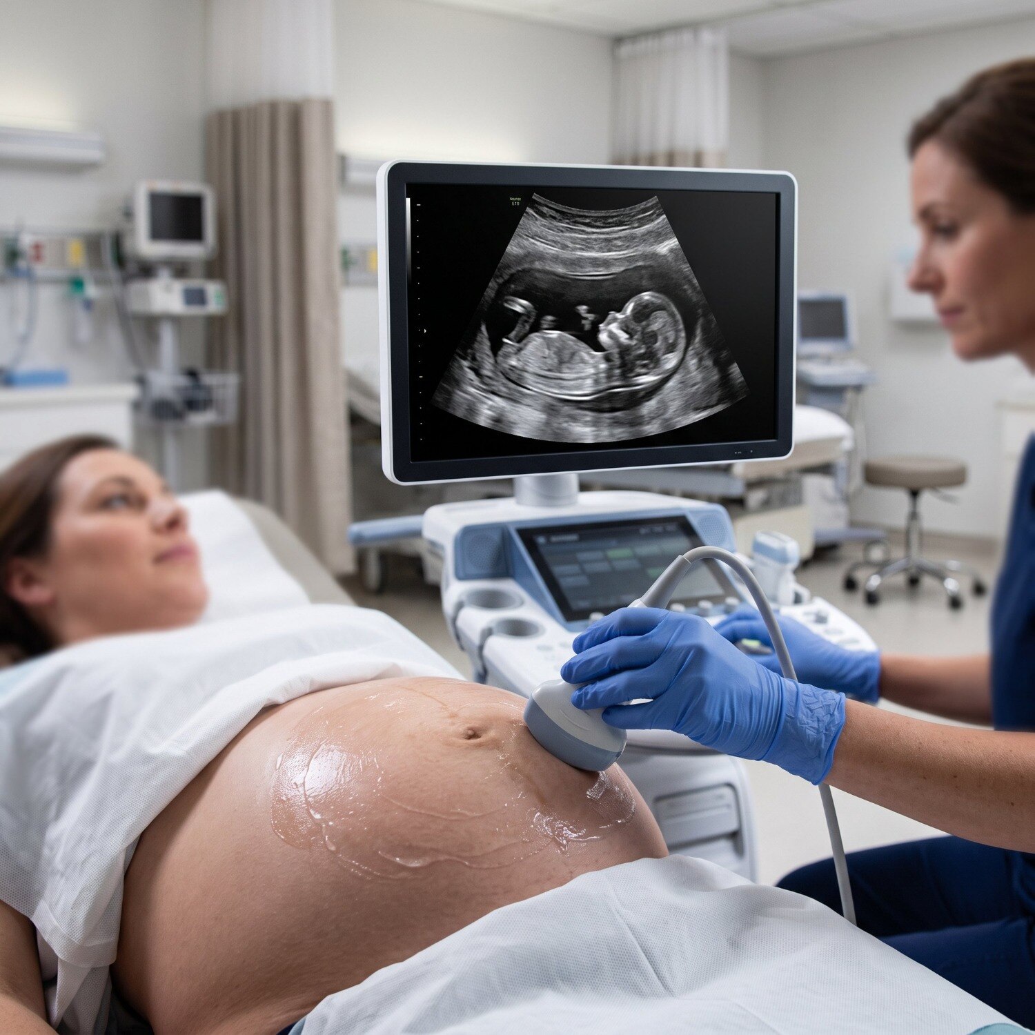
During an abdominal examination, gel is applied to your lower abdomen and the probe captures images. For a transvaginal examination, a covered probe is inserted gently into the vagina; this may give closer views of the cervix, uterus, or early pregnancy and does not mean the pregnancy is abnormal.
The sonologist records required images and measurements, reviews them clinically, and prepares a report.
The scan itself often takes about 15–30 minutes. Complex examinations, waiting, image review, and report preparation can make the total visit longer.
- Choose a provider that identifies the indication, gestational age, findings, limitations, and follow-up advice in the report.
- For ultrasonography for pregnancy in Navi Mumbai, reject any centre that promises fetal-sex disclosure or uses coded language: the Pre-Conception and Pre-Natal Diagnostic Techniques law prohibits determining or communicating fetal sex.
- Medically indicated scans use prudent exposure and are considered safe when performed by trained practitioners. Prolonged non-medical 3D or 4D keepsake scanning should not replace diagnostic care.
| Provider option | What to verify | Best use |
|---|---|---|
| Hospital or diagnostic centre | Qualified practitioner and signed report | Planned or complex scans |
| Ultrasound scan gynecologist Airoli referral | Obstetrician receives the report | Findings needing clinical follow-up |
Take the report to your obstetrician, not to an online calculator. Dr. Namrata Kshatriya-Obstetrics & Gynecology can connect scan findings with maternity or gynecological care when follow-up is needed.
Related services
Antenatal Check Up Antenatal check-ups, also known as prenatal visits, are an essential component of maternity care. View service → |
Ultra Sonography Ultrasound, also known as ultrasonography, is a medical imaging technique that uses high-frequency sound waves to produce real-time images of the... View service → |
Frequently asked questions
When is an early pregnancy ultrasound needed?
An early scan is needed when dates are uncertain, bleeding or pelvic or abdominal pain occurs, or examination raises concern about ectopic pregnancy or miscarriage. It can also follow a previous pregnancy complication or provide a clinically indicated first-trimester assessment.
When is the nuchal translucency scan performed?
The nuchal translucency scan is performed between 11 weeks and 13 weeks 6 days of pregnancy. It measures the fluid at the back of the fetal neck as part of early screening.
What does the 18–22 week anatomy scan check?
The anatomy scan checks fetal organs, the placenta and amniotic fluid, along with overall anatomical development.
What do third-trimester pregnancy scans assess?
Third-trimester scans assess fetal growth, amniotic fluid, Doppler blood flow, movement and presentation, such as whether the baby is head-down.
How should you prepare for a pregnancy ultrasound in Navi Mumbai?
Take your referral or previous scan reports, confirm the gestational age and follow the diagnostic centre’s instructions about food or bladder filling. Choose a centre that provides a written report and images for your obstetrician.
Keywords